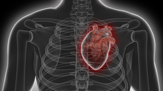
Cardiac catheterization techniques have become indispensable tools in invasive cardiology, enabling physicians to diagnose and treat heart conditions precisely. By guiding a slender catheter through blood vessels directly into the heart, clinicians obtain real-time hemodynamic data and perform targeted interventions. Because these vascular catheterization techniques minimize surgical trauma, patients benefit from faster recoveries and lower complication rates. This educational overview examines the history, imaging innovations, material advancements, and patient-centered practices that define modern cardiac catheterization techniques.
Evolution of Cardiac Catheterization Techniques
The journey of cardiac catheterization techniques began in the early 20th century when researchers first inserted simple tubes into vascular chambers to measure pressure. Yet the groundbreaking work of André Cournand and Dickinson Richards in the 1950s standardized invasive hemodynamic assessments, earning them a Nobel Prize. Over time, these techniques advanced beyond diagnostics: interventional cardiologists introduced balloon angioplasty in the late 1970s, using cardiac catheterization techniques to open narrowed arteries without open surgery.
Since then, operators have refined catheter manipulation strategies and adopted fractional flow reserve measurements to determine which blockages truly impede blood flow. Additionally, structured training—including high-fidelity simulators—prepares new clinicians in essential vascular catheterization techniques before they perform live procedures. Such rigorous preparation has enhanced procedural safety and expanded the applications of cardiac catheterization techniques to include valvular repair and electrophysiology studies.
Imaging Innovations in Cardiac Catheterization Techniques
Advances in imaging have elevated cardiac catheterization techniques by improving procedural accuracy. While fluoroscopy remains the primary modality—offering live X-ray views of catheter position—intracoronary imaging tools like intravascular ultrasound and optical coherence tomography now visualize plaque morphology and vessel wall integrity in exquisite detail. These enhancements allow interventionalists to size stents correctly and confirm optimal device deployment.
Moreover, three-dimensional rotational angiography reconstructs complex anatomies, aiding interventions such as transcatheter valve replacement. Fusion imaging systems merge pre-procedure CT or MRI datasets with live fluoroscopy, guiding operators through challenging anatomies and reducing both radiation exposure and contrast usage. By leveraging these sophisticated cardiac catheterization techniques, care teams can perform complex procedures more safely and efficiently.
Material Advancements in Cardiac Catheterization Techniques
The evolution of catheter materials is at the heart of modern vascular catheterization techniques. Early devices composed of nylon and polyethylene lacked the finesse needed for tortuous vessels. Today’s catheters utilize biocompatible polymers reinforced with braided metallic cores, delivering an optimal balance of flexibility and pushability. Hydrophilic coatings on catheter surfaces reduce friction, allowing smoother navigation through stenotic or calcified vessels.
In therapeutic applications, sensor-tipped catheters provide instantaneous pressure and flow readings without device exchanges, streamlining the procedure. Furthermore, drug-eluting balloons and bioresorbable scaffolds represent the next frontier of vascular catheterization techniques by delivering localized pharmacotherapy and then dissolving, thereby reducing the long-term presence of foreign material within arteries. These innovations make interventions safer and more patient-friendly.
Patient-Centered Practices
Adopting patient-centered approaches enhances the effectiveness of vascular catheterization techniques from consultation through recovery. Prior to intervention, a multidisciplinary heart team reviews diagnostic images, clinical histories, and comorbidities to tailor procedural strategies. During the procedure, fractional flow reserve measurements guide decisions on which lesions require treatment, ensuring that only hemodynamically significant blockages are addressed. Intraprocedural echocardiography—transesophageal or intracardiac—confirms device placement and assesses immediate functional outcomes.
Post-procedure, standardized recovery pathways emphasize early ambulation and patient education about lifestyle modifications, medication adherence, and symptom monitoring. Scheduled noninvasive imaging, such as CT angiography or stress tests, detects any restenosis or device-related issues before symptoms arise. In this way, integrating meticulous vascular catheterization techniques and patient engagement leads to better long-term cardiovascular health.
As innovations continue to propel invasive cardiology forward, mastering cardiac catheterization techniques remains essential for clinicians committed to improving patient outcomes. By blending historical insights, cutting-edge imaging, advanced materials, and patient-centered care, today’s cardiology teams can deliver safer, more precise interventions that set new standards in vascular treatment.